In this article, we’ll explore the essential aspects of pigmented basal cell carcinoma (BCC), covering its symptoms, diagnosis, treatment options, prognosis, and prevention strategies.
Symptoms and Signs of Pigmented Basal Cell Carcinoma
Recognizing pigmented basal cell carcinoma (BCC) symptoms and signs is crucial for early detection and prompt treatment. Here’s what to look out for:
- Changes in Pigmentation: Pigmented BCC often presents as a brown, blue, or black lesion on the skin, which may resemble a mole or melanoma. Keep an eye on any new or existing pigmented lesions that exhibit irregular borders, asymmetry, or variations in color.
- Persistent Bumps or Nodules: Unlike typical moles or freckles, pigmented BCC lesions may appear as raised, shiny, or translucent bumps on the skin. These nodules can vary in size and may bleed or develop crusts over time.
- Ulceration or Crusting: Some pigmented BCCs may ulcerate or develop crusts on the surface, leading to persistent sores that do not heal. If you notice any non-healing wounds or lesions on your skin, especially in sun-exposed areas, it’s essential to have them evaluated by a dermatologist.
- Slow-Growing Lesions: Pigmented BCCs often grow slowly over time, gradually increasing in size and becoming more noticeable. Pay attention to any changes in the appearance or texture of pigmented lesions, as well as any associated symptoms such as itching or tenderness.
- Location on the Body: While pigmented BCCs can occur anywhere on the body, they are more commonly found on sun-exposed areas such as the face, neck, scalp, and upper torso. Be particularly vigilant about monitoring pigmented lesions in these regions, especially if you have a history of sun exposure or sunburns.
- Associated Symptoms: Pigmented BCCs may cause symptoms such as pain, itching, or bleeding in some cases, especially if the lesion becomes irritated or traumatized. If you experience any unusual sensations or discomfort associated with a pigmented lesion, seek medical attention promptly.
- Lymph Node Enlargement: In rare instances, pigmented BCCs may spread to nearby lymph nodes, causing them to enlarge or become tender to the touch. If you notice any unusual lumps or bumps in your lymph nodes, particularly in conjunction with suspicious skin lesions, it’s essential to consult a healthcare professional for further evaluation.
Recognizing these symptoms and signs of pigmented basal cell carcinoma can facilitate early diagnosis and timely intervention, leading to improved outcomes and reduced risk of complications.
Diagnosis and Detection of Pigmented Basal Cell Carcinoma
Diagnosing and detecting pigmented basal cell carcinoma (BCC) relies on a combination of clinical examination, imaging techniques, and biopsy procedures. Here’s what you need to know about the diagnosis and detection of pigmented BCC:
Clinical Examination
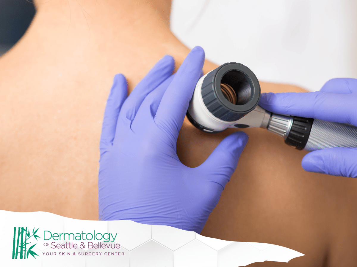
Dermatologists typically begin the diagnostic process by conducting a thorough clinical examination of the skin. They examine any suspicious lesions, including those that are pigmented or have irregular borders, using a dermatoscope—a handheld device that provides magnified views of the skin’s surface.
Pigmented BCC lesions may appear as dark or multi-colored patches, often resembling melanoma, making clinical examination crucial for initial detection.
Dermoscopy
Dermoscopy is a non-invasive imaging technique used to examine pigmented skin lesions in greater detail. Dermoscopy allows dermatologists to visualize skin lesions’ internal structures and pigment patterns, aiding in the differentiation between benign and malignant lesions.
In pigmented BCC, dermoscopy may reveal characteristic features such as blue-gray globules, ulceration, or arborizing vessels.
Biopsy
A skin biopsy is the gold standard for diagnosing pigmented BCC. It involves removing a small sample of skin tissue for microscopic examination by a pathologist. There are several types of skin biopsies, including shave biopsy, punch biopsy, and excisional biopsy, each suited to different lesion types and locations.
During a biopsy, a local anesthetic is typically administered to numb the area before the procedure. The biopsy sample is then examined under a microscope to confirm the presence of cancerous cells and determine the subtype of BCC.
Histopathological Examination
Following a skin biopsy, the excised tissue sample is processed, embedded in paraffin wax, and sliced into thin sections. These sections are then stained with special dyes and examined under a microscope by a pathologist.
The histopathological examination allows for the identification of characteristic features of pigmented BCC, such as nests or islands of basaloid cells with melanin pigment deposition within the tumor tissue.
Differential Diagnosis
Pigmented BCC can mimic other pigmented skin lesions, including melanoma, seborrheic keratosis, and pigmented actinic keratosis. Differential diagnosis involves distinguishing pigmented BCC from these other conditions based on clinical and histopathological features.
Dermatologists rely on their expertise and diagnostic tools, such as dermoscopy, to accurately differentiate between benign and malignant lesions.
Early detection and accurate diagnosis of pigmented basal cell carcinoma are crucial for initiating timely treatment and preventing disease progression. If you notice any suspicious pigmented lesions on your skin, such as dark or multi-colored patches that are changing in size or shape, it’s essential to seek evaluation by a dermatologist promptly.
Key Differences Between Pigmented Basal Cell Carcinoma and Other Skin Cancers

Differentiating between pigmented basal cell carcinoma (BCC) and other types of skin cancer is essential for accurate diagnosis and appropriate treatment. Here are the key differences between pigmented BCC and other common skin cancers:
Basal Cell Carcinoma (BCC) vs. Squamous Cell Carcinoma (SCC)
- BCC originates from the basal cells in the outer layer of the skin, whereas Squamous Cell Carcinoma arises from the squamous cells.
- Pigmented BCC typically appears as dark or multi-colored patches with characteristic features such as blue-gray globules or arborizing vessels, whereas SCC may present as red, scaly, or wart-like growths.
- BCC tends to grow slowly and is less likely to metastasize (spread) compared to SCC, which can be more aggressive and have a higher risk of metastasis.
Pigmented Basal Cell Carcinoma (BCC) vs. Melanoma
- Pigmented BCC arises from basal cells in the skin, while melanoma originates from melanocytes, the pigment-producing cells.
- Pigmented BCC lesions often have irregular borders and may resemble melanoma clinically, but they typically lack the characteristic features of melanoma, such as asymmetry, irregular borders, color variation, and a diameter larger than 6 millimeters (the size of a pencil eraser).
- Unlike melanoma, pigmented BCC is less likely to involve the lymph nodes or metastasize to distant organs.
Pigmented Basal Cell Carcinoma (BCC) vs. Other Benign Lesions
- Pigmented BCC may resemble other benign pigmented lesions, such as seborrheic keratosis or pigmented actinic keratosis, but it often exhibits specific features suggestive of malignancy, such as ulceration, bleeding, or a history of rapid growth.
- Dermatologists use various diagnostic tools, including dermoscopy and skin biopsy, to accurately differentiate between pigmented BCC and benign lesions.
Risk Factors and Sun Exposure:
- Both pigmented BCC and other types of skin cancer are associated with cumulative sun exposure and UV radiation exposure, but they may differ in their risk factors and predisposing conditions.
- Pigmented BCC is commonly found on sun-exposed areas of the head and neck, whereas other skin cancers, such as SCC and melanoma, may occur on other parts of the body or in areas not typically exposed to the sun.
Understanding these key differences between pigmented basal cell carcinoma and other skin cancers is essential for healthcare providers to make an accurate diagnosis and develop an appropriate treatment plan tailored to the individual patient’s needs.
Treatment Options for Pigmented Basal Cell Carcinoma
When it comes to treating pigmented basal cell carcinoma (BCC), healthcare providers consider various factors, including the size, location, and extent of the lesion, as well as the patient’s overall health and preferences. Here are some common treatment options for pigmented BCC:
Surgical Excision
- Surgical excision involves removing the tumor along with a margin of healthy skin tissue to ensure the complete removal of cancerous cells. This method is often preferred for smaller pigmented BCCs or those located in cosmetically sensitive areas.
- Mohs micrographic surgery, a specialized technique that involves removing thin layers of tissue and examining them under a microscope, may be used for larger or recurrent pigmented BCCs to ensure precise removal while sparing healthy tissue.
Electrodesiccation and Curettage (ED&C)
- ED&C is a common outpatient procedure used for superficial pigmented BCCs. It involves scraping away the tumor with a curette (a sharp, spoon-shaped instrument) and then cauterizing the area with an electric needle to destroy any remaining cancer cells.
- While ED&C is effective for superficial lesions, it may not be suitable for deeper or more aggressive pigmented BCCs.
Cryotherapy
- Cryotherapy, or freezing, involves applying liquid nitrogen to the pigmented BCC lesion to destroy cancerous cells. This method is often used for small, superficial lesions and is generally well-tolerated by patients.
- However, cryotherapy may not be as effective for larger or deeper pigmented BCCs and may require multiple treatment sessions for complete clearance.
Topical Therapies
- Certain topical medications, such as imiquimod cream or 5-fluorouracil (5-FU) cream, may be prescribed for treating superficial pigmented BCCs, especially in patients who are not candidates for surgery or other invasive procedures.
- These medications work by stimulating the body’s immune response to target and destroy cancer cells over time. However, they may cause skin irritation or other side effects.
Radiation Therapy
- Radiation therapy may be recommended for pigmented BCCs that are difficult to treat surgically or located in sensitive areas where surgery may not be feasible.
- External beam radiation therapy delivers targeted radiation to the tumor while minimizing damage to surrounding healthy tissue. It may be used as primary treatment or as adjuvant therapy after surgery.
Photodynamic Therapy (PDT)
- PDT involves applying a photosensitizing agent to the pigmented BCC lesion and then exposing it to a specific wavelength of light to activate the medication and destroy cancer cells.
- PDT is often used for superficial or thin-pigmented BCCs and may be an option for patients who prefer a non-invasive treatment approach.
Targeted Therapy and Immunotherapy
- For advanced or metastatic pigmented BCCs that cannot be treated effectively with surgery or other standard therapies, targeted therapy or immunotherapy drugs may be considered.
- These medications target specific molecular pathways involved in cancer growth and proliferation or boost the body’s immune system to recognize and attack cancer cells.
Overall, the choice of treatment for pigmented basal cell carcinoma depends on various factors, and patients should discuss their options with a dermatologist or oncologist to determine the most appropriate course of action based on their individual needs and circumstances.
Final Thoughts
In summary, while this information offers valuable insights into pigmented basal cell carcinoma, seeking personalized medical advice from healthcare professionals for accurate diagnosis and treatment is essential.
Early detection and intervention are crucial for managing this condition effectively and promoting optimal skin health.


