Thinking Outside the Box

a graphic of a tumor on a microscopic level

Quick Summary

  • An internal medicine physician referred a man in his early 60s with expanding forehead redness that was minimally itchy and mainly a cosmetic concern.
  • Two prior dermatology visits resulted in a corticosteroid prescription and dismissal, but the lesion blanched on pressure and had a brownish border that raised concern.
  • The author performed two punch biopsies despite the patient’s cosmetic worries and suspected angiosarcoma based on similar cases seen in residency.
  • Pathology with special testing confirmed angiosarcoma, noted as having a poor prognosis and about a 50% recurrence rate even after treatment.
  • The patient was told he would need coordinated care with radiation oncology, medical oncology, and surgery and was referred to the University of Washington; he and the referring doctor are in-laws expecting their first grandchild.

A nice internal medicine physician, originally Canadian, refers patients to me often from afar. I am very honored and humbled at the same time. Recently, he referred a patient in his early 60s. This patient had seen two dermatologists prior, in addition to his internal medicine physician, one who prescribed a corticosteroid and the other blew him off.

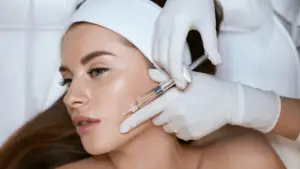
He complained about redness in his forehead, it was not very itchy. It mostly bothered him cosmetically and he wanted laser treatment to remove it.
In a seated position, I examined him from multiple angles. I pressed on it and it blanched. There was no pain whatsoever. It was growing and it had an interesting brownish border and looked like simple chronic sun damage.
Something about this struck me and I recalled photographs that looked almost identical to the gentleman that was sitting in front of me from residency over 20 years ago.

I told him I wanted to make sure he did not have a very rare disease and I needed to do not one but two punch biopsies, even if it left a scar on his face. Mind you, the patient was very cosmetically conscious and the furthest thing from his mind was to add more scars to an area that was already very reddened. To avoid cosmetic damage, he would pay good money to expunge!

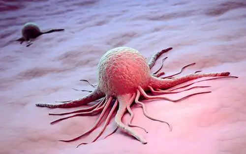
I put down one and only one diagnosis: Angiosarcoma.
About 10 days later, I got the pathology report and after special testing the diagnosis is: angiosarcoma. The prognosis is very poor and there’s a 50% recurrence rate even after all measures taken.

I spoke to the patient as soon as I possibly could to explain that he will need a team of physicians including the radiation oncologist, a medical oncologist, and a surgeon to address this issue.

I referred him to the University of Washington.

The ironic twist in the story is that he and the referring doctor are in-laws, expecting their first grandchild.

FAQ - Frequently Asked Questions

What is angiosarcoma, and why can it look like “just redness” on the forehead?
Angiosarcoma is a rare cancer that arises from cells lining blood or lymphatic vessels. On the face and scalp it can present as a slowly enlarging red or violaceous patch that blanches with pressure, which makes it easy to mistake for sun damage, rosacea, bruising, or dermatitis. It may not itch or hurt, so people often seek care for cosmetic reasons rather than symptoms. Subtle clues like progressive growth, irregular borders, and an unusual color pattern raise concern.
If a red patch is enlarging, changing color, developing a brownish or purplish border, bleeding, or not responding to standard treatments, it deserves a medical evaluation before any cosmetic procedure. A lesion that blanches and continues to spread over weeks to months is also a reason to pause. Cosmetic laser treatment can delay diagnosis and may complicate later assessment. When in doubt, a dermatologist should examine it in good lighting and from multiple angles.
Angiosarcoma can be patchy, and a single small biopsy may miss diagnostic areas. Taking two samples from different parts of the lesion improves the chance of catching the characteristic changes and allows for special stains that confirm the diagnosis. Even when scarring is a concern, biopsy is the safest step because it guides timely, appropriate treatment. Your clinician will usually choose biopsy sites and closure methods to minimize cosmetic impact.
Care is usually coordinated by a multidisciplinary team, often including a surgical oncologist or head-and-neck surgeon, a radiation oncologist, and a medical oncologist. Next steps commonly include staging tests to look for spread and planning treatment that may combine surgery and radiation, with chemotherapy or other systemic therapy considered in some cases. Follow-up is close because recurrence is a known risk with this cancer. The exact plan depends on tumor size, depth, location, and whether there is any evidence of metastasis.
If the redness is new, growing, or atypical, a biopsy should come before laser—diagnosis is the decision point that determines what’s safe. A biopsy visit is often in the range of about $200–$600 in many U.S. practices, but total cost varies with insurance, pathology fees, and whether special stains are needed. Cosmetic laser pricing varies widely based on device type and number of sessions, and it doesn’t address an underlying malignancy if one is present. If your clinician recommends biopsy, it’s because the risk of missing something serious outweighs the risk of a small scar.

Share:

Table of Contents

More Posts

Send Us A Message