Freezing dark spots is a common in-office option when a clinician wants to remove or lighten a superficial spot without cutting the skin. Cryotherapy uses controlled cold, usually applied in seconds, to damage targeted pigment so the body can shed it during healing. This article explains what dark spots are, why they appear, and which types may respond to freezing skin spots with cryotherapy. You will also learn what happens during a typical appointment, what discomfort can feel like, and how to plan for downtime, especially on the face. Finally, we compare cryotherapy for dark spots with other non-invasive skin treatmetns so you can discuss options confidently.
- Cryotherapy targets certain superficial pigmented lesions by controlled freezing
- Healing often includes temporary redness, swelling, and a scab or crust
- Liquid nitrogen for age spots is commonly used, but results depend on spot type and depth
- Aftercare affects irritation and how well color blends during recovery
- Some dark spots respond better to topical agents, lasers, or chemical peels
Introduction to Cryotherapy
Cryotherapy uses extreme cold to destroy unwanted tissue. In dermatology, liquid nitrogen is applied for a short, controlled time to freeze a small area while limiting impact on surrounding skin.
Because it is quick and usually does not involve incisions or stitches, cryotherapy is often grouped with non-invasive skin treatmetns. It may be used for select superficial lesions, including certain pigmented spots, when a clinician determines the lesion is appropriate.
What are Dark Spots?
Dark spots are areas that look darker than surrounding skin due to extra pigment (usually melanin) or pigment left after inflammation. They often appear on sun-exposed skin like the face, hands, and chest, but can also form where skin was injured or irritated.
Treatment depends on the cause and pigment depth. Freezing skin spots tends to work best when pigment is superficial and the spot is discrete and well-defined rather than spread across a larger area.
Causes of Dark Spots
Sun exposure is a leading cause. UV light stimulates pigment-producing cells, which can create uneven tone over time. Hormones, genetics, and aging can contribute, especially in frequently exposed areas.
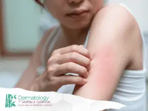
Post-inflammatory hyperpigmentation is another cause, where skin darkens after acne, a rash, an insect bite, or irritation from products or friction. Because pigment is often broader and less defined, topical brighteners, gentle peels, or other non-invasive skin treatmetns are often better first options than freezing.
Types of Dark Spots
Common types include sun spots (age spots), freckles, melasma, and post-inflammatory hyperpigmentation. Some “spots” are benign growths such as seborrheic keratoses, which can be tan to brown and slightly raised.
Cryotherapy for dark spots works best when a lesion can be treated as a specific target, such as certain age spots or stuck-on benign growths. Diffuse conditions like melasma usually need a longer-term plan focused on sun protection, topical therapy, and trigger control rather than spot-freezing.
Cryotherapy for Dark Spots
Cryotherapy for dark spots is usually considered after a clinician confirms the spot appears benign and suitable for freezing. It is often chosen for speed and convenience, especially for a single spot or small cluster.
Results vary by diagnosis and depth. Some spots lighten significantly; others fade partially or need a repeat session. There is also a risk of temporary or lasting lightening in the treated area, especially in deeper skin tones, so expectations and risks should be reviewed first.
What is Cryotherapy?
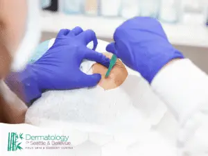
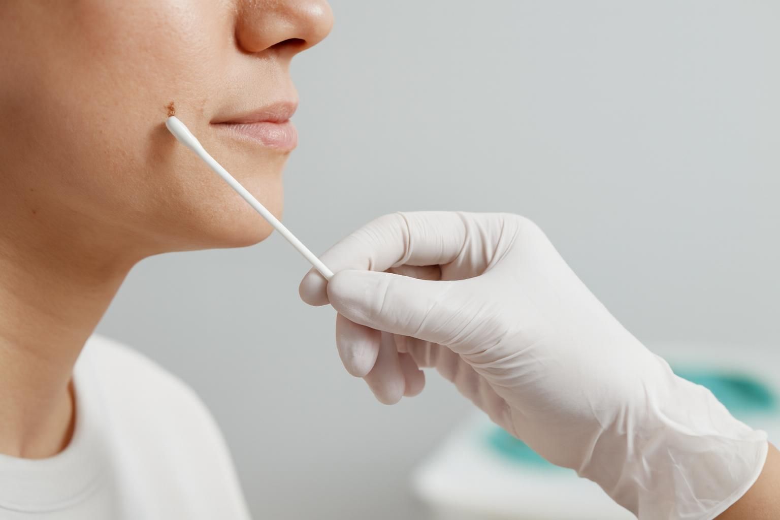
In dermatology, cryotherapy usually means applying liquid nitrogen with a spray device or a cotton-tipped applicator. The clinician performs a controlled freeze, sometimes once and sometimes in a freeze-thaw cycle depending on the lesion.
Visits are brief and do not require stitches. Even so, it is a medical procedure, and the key step is identifying what the spot is, since some lesions should not be frozen and may need a different evaluation.
How Cryotherapy Works
Freezing forms ice crystals inside cells, disrupting membranes and damaging targeted tissue. As the area thaws, blood-flow changes and inflammation add to the injury. Over days, the body clears damaged cells and the surface layer sheds.
For pigmented lesions, the goal is to disrupt pigment-containing cells while minimizing surrounding injury. Clinicians adjust freeze time and intensity based on depth and location. Too little may not treat the spot; too much can increase blistering, scabbing, and uneven pigmentation.
Liquid Nitrogen for Age Spots
Liquid nitrogen for age spots is a common reason people request cryotherapy. Age spots are linked to cumulative sun exposure and often appear as well-defined brown patches on the face and hands. When pigment is superficial and borders are clear, freezing can be an efficient option.
Not every brown spot is an age spot, and some lookalikes need different care. An in-person exam helps confirm the diagnosis and whether cryotherapy fits your skin type, the spot’s location, and your cosmetic goals.
Effectiveness of Liquid Nitrogen
Effectiveness depends on pigment depth and your healing response. Many people see the spot darken briefly, then crust and lift off, revealing lighter skin. Sometimes a faint shadow remains, and a second session or another treatment may be needed.
Downsides can include blistering, scabbing, redness, and pigment changes that last longer than expected. Technique and aftercare may reduce irritation, but they cannot eliminate the risk of uneven tone, especially if the area gets sun during healing.
Comparison with Other Treatments
Cryotherapy is usually faster than topical routines and more targeted than chemical peels, which is helpful for isolated lesions rather than widespread discoloration.
Compared with lasers and light-based devices, cryotherapy can be simpler for single spots, but it is often less precise for blending tone across a larger patch. Topical brighteners and retinoid-like ingredients can support gradual overall improvement, while in-office modalities are often chosen for stubborn, well-defined lesions.
What to Expect During Treatment

A typical visit starts with confirming the spot and reviewing the plan: expected healing, the possibility of pigment change, and how many spots will be treated. Photos may be taken. The area is cleaned, and nearby skin may be protected.
The freeze is brief. The clinician may repeat it, and a follow-up may be needed if the lesion does not respond fully. On the face, clinicians often balance results with cosmetic healing and may use a more conservative freeze.
Procedure Overview
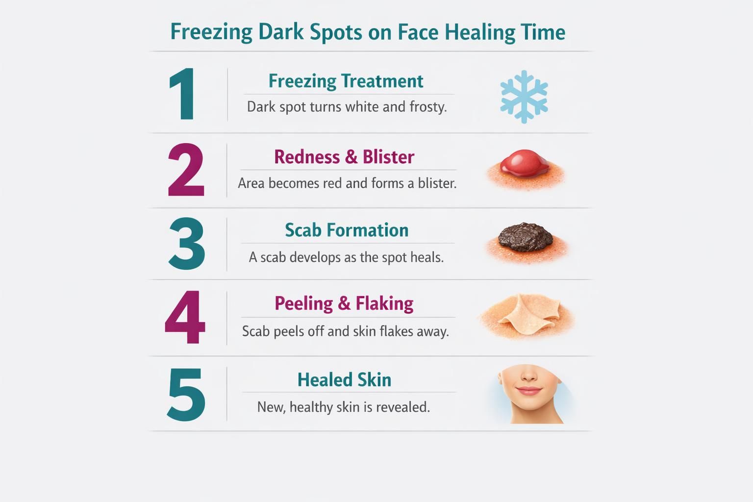
Liquid nitrogen is applied with a spray or applicator for a short interval to freeze the lesion and a small margin. The skin may turn white or frosty during the freeze, then red as it thaws. Some lesions are treated with one freeze; others with a second controlled cycle.
Afterward, the area may sting or feel warm. Mild swelling can develop within hours to a day. A blister may form, especially on thinner skin. Over the next several days, the area can darken, crust, and peel as new skin forms.
Pain Management Options
Discomfort is often a sharp cold sting that fades quickly, but it varies by person and location. The face and hands can feel more intense, and larger areas may stay sore longer.
Pain management is usually simple:
- A clean cold compress afterward if recommended
- Over-the-counter pain relief if appropriate for you
- Avoiding friction from masks, hats, or tight collars for the first day or two
If you are worried about discomfort, ask what sensations are typical for your lesion and whether numbing is an option.
Freezing Dark Spots on Face Healing Time
Freezing dark spots on face healing time is important to plan for because the face is highly visible and often shows redness or crusting during recovery. Many people return to normal activities right away, but may prefer to schedule treatment when a few visible days are acceptable.
Healing depends on spot size, freeze intensity, and individual skin response. Sun protection and avoiding irritation are especially important, since UV exposure can worsen discoloration while skin is healing.
Recovery Process
Right after treatment, redness and a stinging or burning feeling are common and usually settle. Swelling may occur within the first day. A blister can form, or the area may dry, darken, and become a crust as treated tissue sheds.
Over the next several days, the crust loosens and lifts away. Underneath, the skin may look pink or lighter at first and then blend gradually. Picking at the crust, rubbing the area, or sun exposure can prolong healing and increase the risk of pigment changes. Follow your clinician’s instructions for cleansing, moisturizing, and protecting the area until the surface has healed.